


Page 232 - Abstract Book KONIKA 18
P. 232
Hemato-Oncology

                                               P-HO-012
                                  Wilms’ Tumor: A Rare Case Report

                                          Claresta, Ratna Setia Asih
                         Depati Hamzah Regional Hospital, Pangkalpinang, Bangka Belitung, Indonesia

                                               Abstract
            Background Wilms tumor is the most common renal tumor in children. It accounted for 6% of all
            malignancies. The tumors often develop quite large before being noticed and most of them are unilateral.
            Ultrasound, computed tomography, magnetic resonance imaging, and biopsy are routinely used to confirm
            the diagnosis. Objective To report a case of Wilms’ tumor. Case A 7-year-old boy was brought to the
            emergency department for abdominal pain and vomiting in the last 4 days before admission. An abdominal
            mass was also noticed in the left side of the abdomen a month ago, this mass was small in the beginning then
            quickly became larger. Physical examination showed hypertension, pale conjunctiva, and a soft abdomen
            with diffuse tenderness over the upper left area. At the upper left side of the abdominal wall was a mass
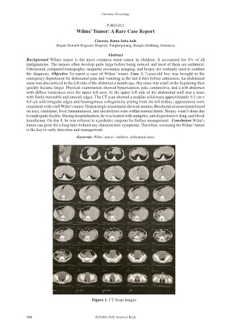
            with freely moveable and smooth edges. The CT scan showed a nodular solid mass approximately 9.5 cm x
            4.8 cm with irregular edges and heterogenous echogenicity arising from the left kidney; appearances were
            consistent with a left Wilm’s tumor. Hematologic assessment showed anemia. Biochemical assessment based
            on urea, creatinine, liver transaminases, and electrolytes were within normal limits. Biopsy wasn’t done due
            to inadequate facility. During hospitalization, he was treated with analgetic, anti-hypertensive drug, and blood
            transfusion. On day 8, he was referred to a pediatric surgeon for further management.  Conclusion Wilm’s
            tumor can grow for a long time without any characteristic symptoms. Therefore, screening for Wilms’ tumor
            is the key to early detection and management.
                                  Keywords: Wilms’ tumor; children; abdominal mass


                                        Figure 1. CT Scan images


            184                           KONIKA XVIII Abstract Book

